

The complex abilities of the hand are part of what makes humans unique. Only humans have the ability to bring our thumbs across the hand to connect with our ring and pinkie fingers. This ability provides us with the dexterity to use tools. It also gives us a forceful grip.
The hand can be considered in four segments:
-
-
- Fingers: Digits that extend from the palm of the hand, the fingers make it possible for humans to grip the smallest of objects.
- Palm: This is the bottom of the body of the hand.
- Back (opisthenar): The back of the hand shows the dorsal venous network, a web of veins.
- Wrist: The connection point between the arm and the hand, the wrist enables hand movements.
-
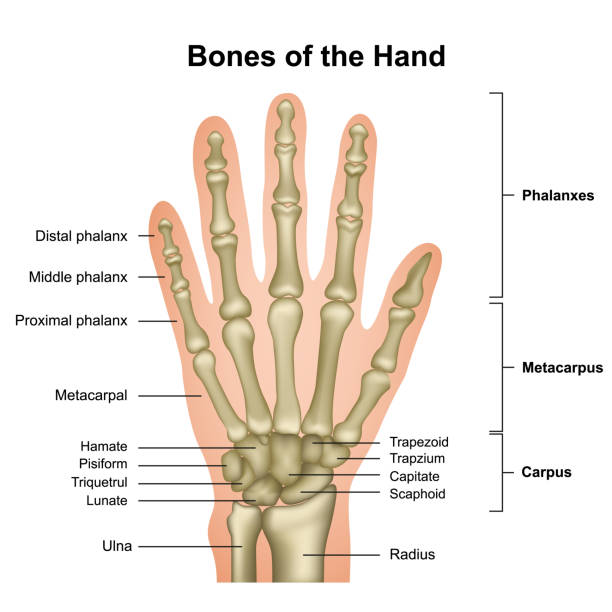
Each hand consists of 19 bones. The palm includes five metacarpals, and each finger except the thumb contains one proximal phalanx, one middle phalanx, and one distal phalanx. The thumb doesn’t have a middle phalanx. Each bone is connected by a series of ligaments.
Each fingertip—distal phalanx and accompanying tissue—contains a fingernail. These structures are made of keratin, a tough protein. Similar types of keratin also make up human hair, the scales and claws of reptiles, and the feathers, claws, and beaks of birds.
The palm of the hand doesn’t contain melanin (skin pigment) or hair follicles. The only other place on the body that lacks both of these is the sole of the foot. These two surfaces also have thicker skin than other places of the body.
The thumb is the first of the hand’s five digits, but it is typically not referred to as a finger. The thumb possesses a unique and wide range of motion not shared by the hand’s other digits. Not only does it bend its knuckle, but the tip of the thumb can touch the tips of the fingers. This range of motion greatly assists in the ability to grasp and hold items.
Anatomically, the thumb is compromised of the metacarpal connected to the trapezium, a carpal bone in the wrist. This first metacarpal connects to the proximal phalanx. This connects to the thumb’s distal phalanx, which is also the tip of the digit. Unlike fingers, the thumb does not possess an intermediate phalanx bone.
The thumb generally receives oxygenated blood by the princeps pollicis artery. The muscles of the thumb all contain the word ‘pollicis,’ and are distinguished by extensor, flexor, oppenens, and abductor designations. This includes longus and brevis descriptions. The first dorsal interosseus also serves the thumb.
The hand is composed of many different bones, muscles, and ligaments that allow for a large amount of movement and dexterity. There are 3 major types of bones in the hand itself, including:
-
-
- Phalanges. The 14 bones are found in the fingers of each hand and also in the toes of each foot. Each finger has 3 phalanges (the distal, middle, and proximal); the thumb only has 2.
- Metacarpal bones. The 5 bones compose the middle part of the hand (Palm).
- Carpal bones. The 8 bones that create the wrist. The 2 rows of carpal bones are connected to 2 bones of the arm–the ulna bone and the radius bone.
-
Numerous muscles, ligaments, tendons, and sheaths can be found within the hand. The muscles are the structures that can contract, allowing movement of the bones in the hand. The ligaments are fibrous tissues that help bind together the joints in the hand. The sheaths are tubular structures that surround part of the fingers. The tendons connect muscles in the arm or hand to the bone to allow movement.
In addition, there are arteries, veins, and nerves within the hand that provide blood flow and sensation to the hand and fingers.
What are knuckles?
A knuckle is a finger joint. The knuckles are where the two phalanges, or finger bones, meet each other and where they meet the metacarpals, or hand bones.
Most people have 14 knuckles in each hand, three on each finger, and two on each thumb.
Complex networks of nerves, muscles, tendons, and ligaments allow the knuckles to move, giving rise to precise finger movements, such as typing, holding utensils, and scratching the head.
Muscles

The muscles of the hand are the skeletal muscles responsible for the movement of the hand and fingers. The muscles of the hand can be subdivided into two groups: the extrinsic and intrinsic muscle groups. The extrinsic muscle groups are the long flexors and extensors. They are called extrinsic because the muscle belly is located on the forearm. The intrinsic group is the smaller muscles located within the hand itself. The muscles of the hand are innervated by the radial, median, and ulnar nerves from the brachial plexus.
The intrinsic muscle groups are the thenar (thumb) and hypothenar (little finger) muscles; the interossei muscles (four dorsally and three volarly) originating between the metacarpal bones; and the lumbrical muscles arising from the deep flexor (and which are special because they have no bony origin) to insert on the dorsal extensor hood mechanism. Palmaris brevis which is a superficial muscle and adductor pollicis are also intrinsic muscles.[2]
Extrinsic group
The fingers have two long flexors, located on the underside of the forearm. They are inserted by tendons to the phalanges of the fingers. The deep flexor attaches to the distal phalanx, and the superficial flexor attaches to the middle phalanx. The flexors allow for the actual bending of the fingers. The thumb has one long flexor and a short flexor in the thenar muscle group. The human thumb also has other muscles in the thenar group (opponens and abductor brevis muscle), moving the thumb in opposition, making grasping possible.
The extensors are located on the back of the forearm and are connected in a more complex way than the flexors to the dorsum of the fingers. The tendons unite with the interosseous and lumbrical muscles to form the extensor hood mechanism. The primary function of the extensors is to straighten out the digits. The thumb has two extensors in the forearm; the tendons of these form the anatomical snuff box. Also, the index finger and the little finger have an extra extensor, used, for instance, for pointing. The extensors are situated within 6 separate compartments.
| Compartment 1 (Most radial) | Compartment 2 | Compartment 3 | Compartment 4 | Compartment 5 | Compartment 6 (Most ulnar) |
|---|---|---|---|---|---|
| Abductor pollicis longus | Extensor carpi radialis longus | Extensor pollicis longus | Extensor indicis | Extensor digiti minimi | Extensor carpi ulnaris |
| Extensor pollicis brevis | Extensor carpi radialis brevis | Extensor digitorum communis |
The first four compartments are located in the grooves present on the dorsum of the inferior side of the radius, while the 5th compartment is in between the radius and ulna. The 6th compartment is in the groove on the dorsum of the inferior side of the ulna.
Nervous System
Three nerves control function in our hands: the median, ulnar, and radial nerves. Each of these nerves is responsible for both sensory and motor function in different parts of the hand.
-
-
- The median nerve. This nerve originates at the shoulder and controls the muscles we need to perform fine precision hand movements and pinching functions. The median nerve is the only nerve that enters the hand through the carpal tunnel; a space formed by the carpal bones of the wrist. This nerve controls sensation in the thumb, index finger, middle finger, and one side of the ring finger.
-
-
-
- The ulnar nerve. The ulnar nerve runs through the arm into the hand and is the largest unprotected nerve in the human body. It connects to the little finger and adjacent side of the ring finger of the hand, providing sensation on the palm side of the hand. The ulnar nerve enables us to grasp objects. It travels along the elbow, between the bone, and overlying skin at the cubital tunnel. When we bump our “funny bone,” the painful sensation we feel comes from impact against this nerve. The ulnar nerve enters the palm of the hand through the Guyon’s canal.
-
-
-
- The radial nerve. This nerve runs through the arm and controls our ability to extend our wrist and control the position of our hand. It also provides sensory feedback from the back of the little finger and the adjacent half of the ring finger.
-
Ailments
Although fully functional hands can accomplish great things, they are susceptible to a number of ailments, including:
-
-
- Arthritis
- Deformities
- Nerve disorders
- Finger clubbing
- Tendinitis
- Carpal tunnel syndrome
- Fractured bones
- Sprains, strains, cuts, and bruises
-
Typically, four main issues cause knuckle pain.
Injury
Injuring any of the structures that make up the knuckles can lead to knuckle pain. Types of knuckle injuries include:
-
-
-
- Strains: including stretched or torn muscles and tendons
- Sprains: stretched or torn ligaments
- Fractured or broken knuckles: can occur when a person punches something or falls on their hand
- Dislocated knuckle: when a phalanx, or finger bone, moves out of its original position
-
-
Learn about the differences between a strain and a sprain here.
Symptoms of a knuckle injury
The symptoms of a knuckle injury vary depending on the type and severity of the damage.
People may feel mild to severe pain in the injured knuckle or knuckles. This can feel like a dull ache or a sharp, stabbing sensation.
The pain may worsen with movement.
Symptoms of a strain or sprain near a knuckle include:
-
-
-
- pain
- swelling
- limited flexibility
- stiffness or reduced range of motion
-
-
Symptoms of a fractured or broken knuckle include:
-
-
-
- bruising
- limited or no range of motion
- numbness or tingling in the affected finger
- visible changes, such as swelling and discoloration
- finger appears bent at an abnormal or extreme angle
-
-
Treatments for knuckle injuries
People can treat sprains, strains, and other mild knuckle injuries with RICE therapy:
-
-
-
-
- Rest: Avoid moving or using the injured knuckle while it heals. Consider immobilizing the knuckle in a splint or brace.
- Ice: Apply an ice pack or cold compress on the injured knuckle. This will help reduce swelling and relieve pain.
- Compression: If the knuckle appears swollen, consider wrapping it with a bandage or buddy tape.
- Elevation: Keep the injured finger raised above the heart. This may help reduce swelling.
-
-
-
A healthcare provider can diagnose a fractured, broken, or dislocated knuckle.
They will reset broken bones or maneuver a dislocated phalanx back into place. They may recommend wearing a brace or splint while the knuckle heals.
Over-the-counter (OTC) pain relievers, such as nonsteroidal anti-inflammatory drugs (NSAIDs), can reduce swelling and pain.
Rheumatoid arthritis
Rheumatoid arthritis (RA) is an autoimmune disease that leads to inflammation in the joints. The Arthritis Foundation state that RA typically affects the joints in the hands, wrists, and knees.
According to an overview article in Medical Principles and PracticeTrusted Source, RA initially causes painful swelling in small joints, such as the knuckles, before progressing to larger joints and organs.
Symptoms of RA in the knuckles include:
-
-
-
-
- tenderness or pain in the knuckles of one or both hands
- knuckles feel stiff, especially in the morning
- pain and stiffness occur in other joints, such as the wrists, knees, or hips
-
-
-
RA can lead to widespread symptoms, such as fatigue and weight loss.
The goals of RA treatment include:
-
-
-
-
- relieving inflammation
- managing other symptoms, such as pain and fatigue
- improving joint function and mobility
- preventing long-term complicationsTrusted Source, such as heart disease, obesity, and reduced quality of life
-
-
-
A healthcare provider may recommend one or more of the following treatments for knuckle pain related to RA:
-
-
-
-
- NSAIDs, immunosuppressants, or corticosteroids to reduce inflammation and pain
- topical pain relievers
- physical therapy to improve joint mobility
- heat therapy to relax the finger muscles and promote joint lubrication
- cold therapy to reduce inflammation and pain
-
-
-
Osteoarthritis
Osteoarthritis (OA) is the most common type of arthritis, affecting more than 32.5 million adultsTrusted Source in the United States.
It is an age-related degenerative disease that involves cartilage loss and bone changes. OA can affect any joint in the body, including the knuckles.
OA most often affects the joints at the tip and middle of the fingers. OA can also cause swelling at the base of the thumb.
Symptoms of OA include:
-
-
-
-
- swelling in multiple knuckles
- deep, aching pain in the affected knuckles
- joint stiffness, especially in the morning
- reduced flexibility and limited range of motion in the affected knuckles
- muscle weakness near the knuckles
- a clicking or popping sound when bending the knuckles
-
-
-
There is no cure for OA. However, combining medication, physical therapy, and lifestyle changes, such as exercise and weight control, can help relieve OA symptoms and prevent long-term complications.
Medications for OA include:
-
-
-
-
- OTC pain relievers, such as acetaminophen and ibuprofen
- Topical creams or gels that contain lidocaine, menthol, or capsaicin
- oral or injectable corticosteroids
- platelet-rich plasma (PRP) injections
-
-
-
Non-drug therapies for OA include:
-
-
-
-
- Physical therapy exercises to strengthen the joints and reduce stiffness
- surgery to remove damaged cartilage, for example
-
-
-
Ganglion cyst
A ganglion cyst is a round, fluid-filled growth that usually develops on the back of the wrist and the base of the fingers. Ganglion cysts can also appear on the knuckle closest to the tip of the finger.
Only about 10% of ganglion cystsTrusted Source form on joints other than the wrist.
A ganglion cyst may feel painful or tender to the touch. The pain only affects the knuckle where the cyst developed.
A ganglion cyst may resolve suddenly without medical treatment. A healthcare provider may also use a needle to drain a ganglion cyst.
A doctor may need to surgically remove a ganglion cyst if it returns or does not respond to other treatments.
Other causes of knuckle pain
Other possible causes of knuckle pain include:
-
-
-
-
- soft tissue growths, such as boils or nodules
- swollen tendons, also known as tendinitis
- gout
- infections
- bone cancer
- multiple sclerosis (MS)
- Raynaud’s phenomenon
- scleroderma
- lupus
- polymyositis
-
-
-
Symptoms of tendonitis in your finger
a lump or bump in or around the tendon. swollen fingers. cracking or snapping feeling when bending your finger. heat or warmth in the affected finger.
Hand tendon repair
If any of the tendons in your hand are damaged, surgery may be needed to repair them. It can also help restore movement in the affected fingers or thumb.
What are tendons?
Tendons are tough cords of tissue that connect muscles to bones. When a group of muscles contract (tighten), the attached tendons will pull on certain bones. This will allow you to make a wide range of movements.
There are 2 groups of tendons in the hand:
-
- extensor tendons – run from the forearm, across the back of your hand to your fingers and thumb, allowing you to straighten your fingers and thumb
- flexor tendons – run from your forearm, through your wrist, and across the palm of your hand, allowing you to bend your fingers
Surgery can often be carried out to repair damage to both these groups of tendons.
When hand tendon repair is needed
Hand tendon repair is carried out when one or more tendons in your hand rupture or are cut, leading to loss of normal hand movements.
If your extensor tendons are damaged, you’ll be unable to straighten one or more fingers. If your flexor tendons are damaged, you’ll be unable to bend one or more fingers. Tendon damage can also cause pain and inflammation (swelling) in your hand.
In some cases, damage to the extensor tendons can be treated without the need for surgery. This may be done using a rigid support called a splint that’s worn around the hand.
Common causes of tendon injuries
How Hand tendons are repaired
Before the cut tendons in your hand are repaired, X-rays of your hand and forearm may be taken. This is to check for:
-
-
-
-
- fragments of glass that may have cut the tendon
- any other damage that needs to be repaired, like a fracture
-
-
-
Tendon repair isn’t usually regarded as emergency surgery. But, it’s generally carried out as quickly as possible after the injury – usually within a few days. This is because the longer the tendons remain ruptured, the more scarring will develop on the end of the tendons. This could reduce the range of your hand movement after surgery.
Depending on the nature of your injury, you may be given antibiotics and a tetanus jab before surgery. This will help to prevent your hand from becoming infected.
Extensor tendon repair
Extensor tendon repair is usually carried out either under a regional or a general anesthetic.
For a regional anesthetic, an injection is used to make part of your body totally numb. For hand surgery, regional anesthetic is injected into the base of the neck or the top of the shoulder to numb the whole arm.
If your tendon is damaged as a result of a wound, the wound will be thoroughly cleaned. An incision may be made in your hand to make the wound larger. The two ends of the ruptured tendon will then be stitched together.
The wound will be closed with stitches. A rigid splint (support to protect your hand) made of plaster will usually be fitted to stop you from moving your hand and damaging the repaired tendons.
Extensor tendon repair surgery can take around 30 minutes to complete if nothing else has been damaged.
Flexor tendon repair
Flexor tendon repair is also usually carried out under either a regional or general anesthetic.
A tourniquet will be wrapped around your upper arm to stop the blood circulating. This is so that bleeding at the wound doesn’t make it difficult to see the relevant structures. A tourniquet is a cord or tight bandage used to constrict (squeeze) the arm and temporarily cut off the blood supply.
The surgeon will then extend the wound, or make an incision if there’s no wound, to locate the damaged tendons. They’ll bring the two ends of the damaged tendon together, before stitching them to each other.
The wound in the hand will be closed with stitches. A rigid plaster splint will usually be applied to protect the repaired tendons.
A simple flexor tendon repair takes 45 to 60 minutes. But, complex surgery for more severe injuries could take much longer
References: healthline, John Hopkins Medicine,  , Wikipedia

